Describe the Method of Radiology Called a Ct Scan
Computed tomography CT scanning also known as especially in the older literature and textbooks computerised axial tomography CAT scanning is a diagnostic imaging procedure that uses x -rays to build cross-sectional images slices of the body. CT scan computerized tomography is a procedure that uses X-rays to scan and take images of cross-sections of parts of the body.

Computed Tomography Ct Or Cat Scan Of The Bones Johns Hopkins Medicine
A radiographic technique in which a radiopaque shows up on X-ray contrast material is injected into a blood vessel for the purpose of identifying its anatomy on an X-ray.
. A computed tomography CT or CAT scan allows doctors to see inside your body. They are sometimes called CAT computer-assisted tomography scanners. CT scanners a type of Xray machine became important for diagnosis within hospitals during the late 20th century.
A CT scanner takes multiple cross-sectional images of the body. CT scan can help diagnose broken bones tumors or lesions in areas of the body blood clots in the brain legs and lung and lung infections or diseases like pneumonia or emphysema. Computed tomography CT scanning also known as especially in the older literature and textbooks computerized axial tomography CAT scanning is a diagnostic imaging procedure that uses x -rays to build cross-sectional images slices of.
The first detailed picture of a living brain was taken by a CT scanner in 1971. CT provides more detail than an X-ray and can better define areas where tissues overlap. Cone beam computed tomography CBCT -acquires all the data in a single sweep of the scanner employing a large cone-shaped x-ray beam matched with a flat panel detector for volume acquisition of data.
For a better look at other areas an intravenous IV contrast will be used. This will provide more detail of your stomach and intestines. During the test you will lie on a table that is attached to the CT scanner which is a large doughnut-shaped machine.
A computed tomography CT scan uses X-rays to make detailed pictures of structures inside of the body. -allows shorter scanning times and. Also called contrast resolution or contrast detectability.
Computed axial tomography CAT scans or CT scans use a series of X-rays plus a computer to produce a cross-sectional image of the inside of the body. The pixels are and external branches obtained from a spiral CT scan. The term computed tomography or CT refers to a computerized x-ray imaging procedure in which a narrow beam of x-rays is aimed at a patient and quickly rotated around the body producing signals that are processed by the machines computer to generate cross-sectional imagesor slicesof the body.
Computed Tomography also called a CAT scan or CT is a non-invasive medical test that uses a series of cross-sectional images to view a bodily organ structure or system. The CT scanner consists of a table which supports the patient and a large ring that rotates 360 degrees around the area being studied taking multiple images from every angle in just seconds. CT is accomplished in three stepsscanning the patient data acquisition processing the data image reconstruction and displaying the image.
Also called high-contrast resolution or detail resolution click to flip Dont know Question Ability of the system to differentiate between objects with similar densities. Then a radiologist looks at the CT images and talks to the doctor to see whats going on inside the person and if. CT has proven so valuable as a medical diagnostic tool that the 1979 Nobel Prize in Medicine was awarded to the inventors.
Computed tomography CT or CAT scan ranks as one of the top five medical developments in the last 50 years according to most medical surveys. If a person feels unwell or has pain doctors might suggest having a CT scan to find out what the problem is. A computerized tomography CT scan also called a computed tomography or CAT scan is a scan that takes very detailed pictures of the inside of the body.
Early CT scanner in use 1980. This technique is used to image arteries in the brain heart kidneys gastrointestinal tract aorta neck carotids chest limbs and pulmonary circuit. CT scans can detect smaller abnormalities than can be found with a conventional X-ray.
The method used to scan is dependent upon the equipment. The first step scanningthe patient is the radiographic portion of the study. If you are having a CT scan of the abdomen or pelvis please call Advanced Radiology at 401866 727-4600 for instructions on preparation since these studies may require a barium drink.
Figure 5b is an The simplest method is a threshold operation applied image of the common carotid arteries and their internal to the original gray values of the images. The CT scanner sends X-rays through the body area being studied. Computed tomography scan Also called a CT or CAT scan - a diagnostic imaging procedure that uses a combination of x-rays and computer technology to produce cross-sectional images often called slices both horizontally and vertically of the body.
A completed CT scan of the chest usually consists of two complete sets of images one showing mediastinal structures in which the lung fields look relatively black and one showing the lung fields in which the mediastinum looks relatively white. Question Ability of a system to resolve as separate forms small objects that are very close together. CT stands for computerized tomography.
Computed tomography CT also known as a computerized axial tomography CAT scan including CT angiography Fluoroscopy including upper GI and barium enema Magnetic resonance imaging MRI and magnetic resonance angiography MRA Mammography Nuclear medicine which includes such tests as a bone scan thyroid scan and thallium cardiac. It uses a combination of X-rays and a computer to create pictures of your organs bones and.

X Ray Computed Tomography An Overview Sciencedirect Topics

Diagnostic Tests X Ray Ct Scan Mri
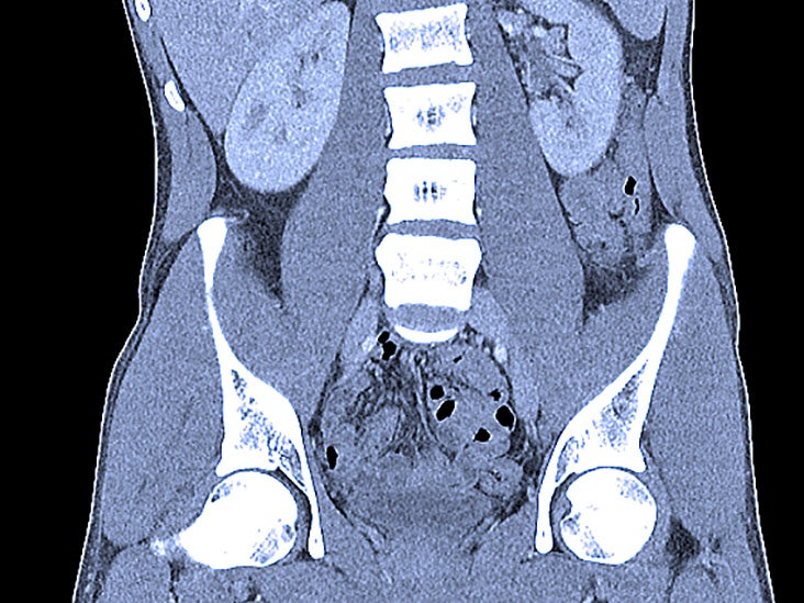
Abdominal Ct Scans Definition Uses Picture And More
No comments for "Describe the Method of Radiology Called a Ct Scan"
Post a Comment